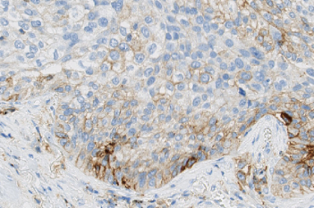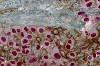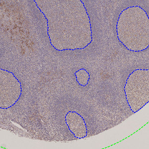
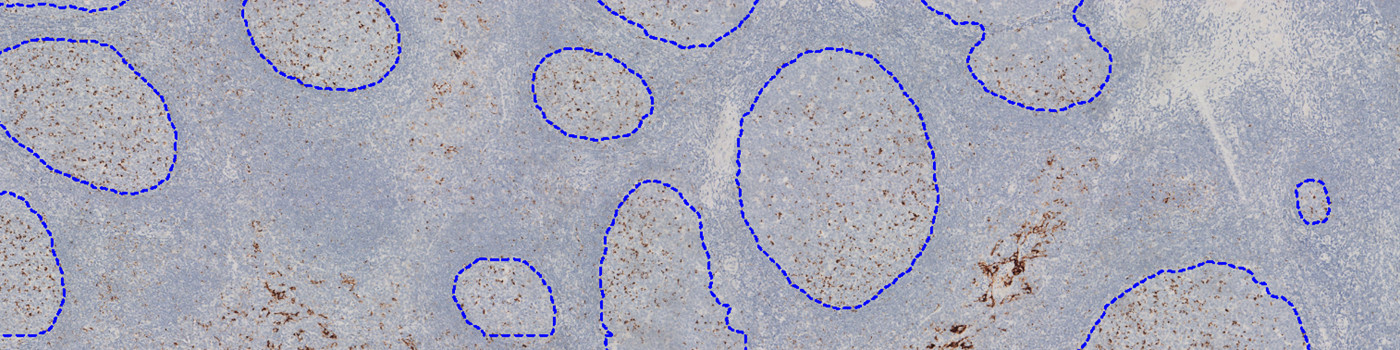
Outlined germinal centers (blue) on TMA core.
#10163
Developed for outlining germinal centers in tonsils
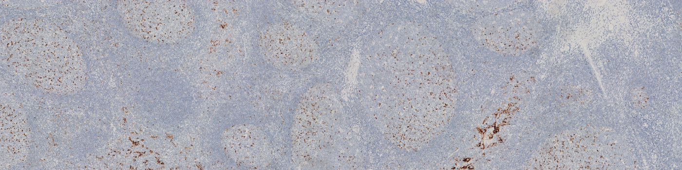
Tonsil tissue may be used as positive/negative controls for PD-L1 staining. Because the PD-L1 expression varies significantly between the tissue compartments, it is of interest to differentiate between tissue regions before evaluating the PD-L1 expression.
This APP automatically outlines germinal centers in tonsils stained for PD-L1 and is intended as an accessory APP to help isolate regions for further analysis.
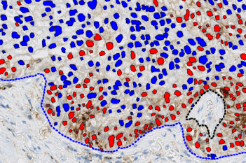
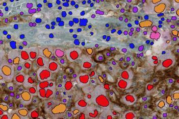
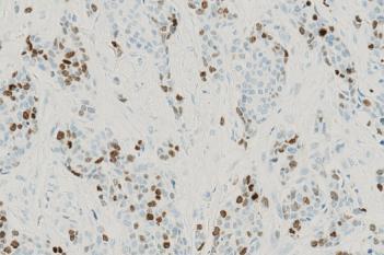
Outlined germinal centers (blue) on TMA core.
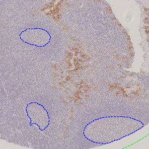
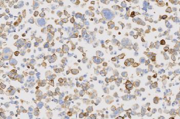
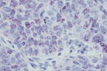
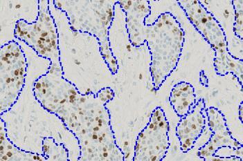
Outlined germinal centers (blue) on TMA core.
Quantitative Output variables
The output variables obtained from this protocol are:
Workflow
Step 1: Load and run the APP 01 Germinal Center Detection
Methods
The APP was developed using the using the DeepLabv3+ neural network available with Author™ AI. The neural network uses a cascade of layers of nonlinear processing units for feature extraction and transformation, with each successive layer using the output from the previous layer as input. DeepLabv3+ uses an encoder-decoder structure with atrous spatial pyramid pooling (ASPP) that is able to encode multi-scale contextual information by probing the incoming features with filters or pooling operations at multiple rates and multiple effective field-of-views. This means that instead of using step-wise upsampling blocks to incorporate features from different levels, this network only needs two upsampling steps, i.e. it is faster to train and analyze than e.g. the U-Net. All of this also means that the decoder module can refine the segmentation results along the object boundaries more precisely. For more information on the network architecture, see [1].
Staining Protocol
There is no staining protocol available.
Additional information
To run the APP, a NVIDIA GPU with minimum 4 GB RAM is required.
Keywords
Tonsil, Germinal Center, PD-L1, Quality Control, AI, Deep Learning
METHODS
References
LITERATURE
1. Chen, L., et. al., Encoder-decoder with atrous separable convolution for semantic image segmentation, Proceedings of the European conference on computer vision (ECCV) 2018, 801-818, DOI