Description
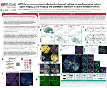
The ability to image tissue microenvironments (TME) at high-plex over an entire slide without requiring multiple staining steps allows for unprecedented insights into tissue architecture and the molecular mechanisms of immune and disease processes. Here we investigate a whole slide tissue section of high-grade colon adenocarcinoma, a hot tumor that contains prominent clusters of PD-L1 expression scattered throughout, using single-step high-plex staining and imaging at single-cell resolution followed by the analysis of single-cell phenotypes, tissue segmentation and spatial proximity and nearest neighbor analysis.
Authors and institutions
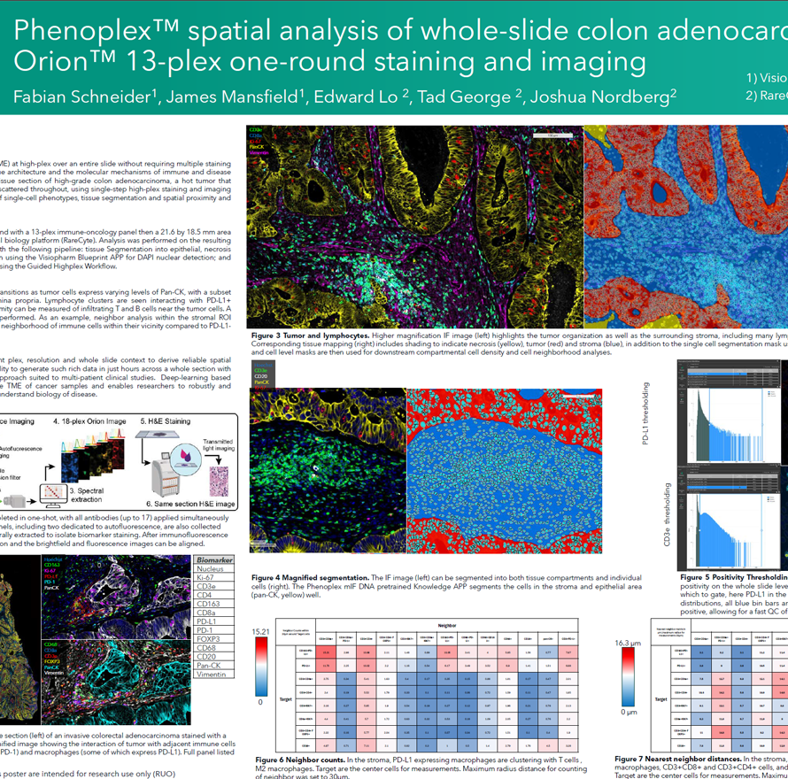
Fabian Schneider1, James Mansfield1, Edward Lo2, Tad George2, Joshua Nordberg2
- Visiopharm A/S, Horsholm, Denmark
- RareCyte, Seattle, WA