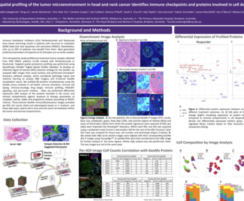
Find out why Dr Rimm’s work suggests that a subset of cells that have lost beta-catenin expression may be associated with non-response. In this seminar recording from USCAP 2020, Dr Rimm explains how you can benefit from exploratory analysis using AI-powered phenotyping.
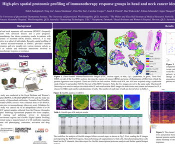
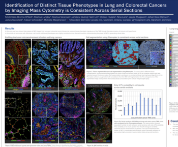
Learn how his group used Visiopharm AI-based software to help them get meaningful results from next-gen imaging technologies like Fluidigm’s IMC.
-
- Methods of measurement –co-localization, compartmentalization, and measurement vs segmentation and counting
-
- How his lab used tSNE plots to show differences between responders and non-responders to trastuzumab

David Rimm, MD, PhD, Professor of Pathology and Medicine (Oncology); Director of Yale Pathology Tissue Services. Yale School of Medicine
David has authored over 400 peer-reviewed papers and holds eight patents. His research lab group focuses on quantitative pathology using the AQUA® technology invented in his lab, and other quantitative methods, including Visiopharm´s phenotyping module.
His projects relate to predicting response to both targeted and immune- therapy in cancer and standardization of those assays for CLIA labs.